44 structure of the heart without labels
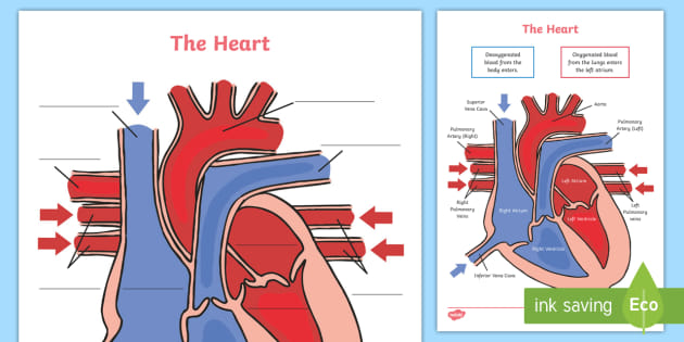
circulatory system worksheet without labels - Google Search | Heart ... Students will color these areas RED. The areas of the heart with LESS oxygen are labeled with a "B". Students will color these areas BLUE. Re-pin and click on this item to learn more! #cardiovascularsystem #humanbody #circulatorysystem #bodysystems #anatomy #heart #healthclass #humanbody #biology #lifescience The Anatomy of the Heart - Quiz 1 - Free Anatomy Quiz The circulatory system - lower body image, with blank labels attached. The circulatory system - a PDF file of the upper and lower body for printing out to use off-line. Describe and explain the function of the circulatory system - The circulatory system consists of the heart, the blood vessels (veins, arteries, and capillaries), and the blood.
Heart anatomy: Structure, valves, coronary vessels | Kenhub The heart is shaped as a quadrangular pyramid, and orientated as if the pyramid has fallen onto one of its sides so that its base faces the posterior thoracic wall, and its apex is pointed toward the anterior thoracic wall.

Structure of the heart without labels
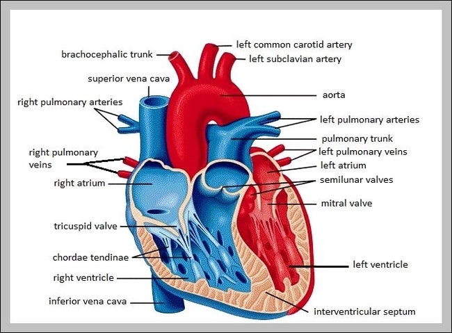
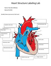
Human Heart Diagram Without Labels - Labelling Worksheet The human heart is a muscle made up of four chambers, these are: Two upper chambers - the left atrium and right atrium Two lower chambers - the left and right ventricles. It's also made up of four valves - these are known as the tricuspid, pulmonary, mitral and aortic valves. A Labeled Diagram of the Human Heart You Really Need to See The human heart, comprises four chambers: right atrium, left atrium, right ventricle and left ventricle. The two upper chambers are called the left and the right atria, and the two lower chambers are known as the left and the right ventricles. The two atria and ventricles are separated from each other by a muscle wall called 'septum'. WebMD - Better information. Better health. The heart is a muscular organ about the size of a fist, located just behind and slightly left of the breastbone. The heart pumps blood through the network of arteries and veins called the...
Structure of the heart without labels. The Heart | Boundless Anatomy and Physiology | | Course Hero The myocardium is the muscle tissue of the heart, composed of cardiac muscle cells called cardiomyocytes that receive nervous stimulation from the sinoatrial (SA) and atrioventricular (AV nodes via the Purkinje fibers. Cardiomyocytes are shorter than skeletal myocytes, and contain fewer nuclei. Cardiac muscle is striated. Human Heart - Diagram and Anatomy of the Heart - Innerbody Because the heart points to the left, about 2/3 of the heart's mass is found on the left side of the body and the other 1/3 is on the right. Anatomy of the Heart Pericardium. The heart sits within a fluid-filled cavity called the pericardial cavity. The walls and lining of the pericardial cavity are a special membrane known as the pericardium. Human Heart - Anatomy, Functions and Facts about Heart Briefly explain the structure of the human heart. The human heart is divided into four chambers, namely two ventricles and two atria. The ventricles are the chambers that pump blood and atrium are the chambers that receive the blood. DNA - Wikipedia The structure of DNA is dynamic along its length, being capable of coiling into tight loops and other shapes. In all species it is composed of two helical chains, bound to each other by hydrogen bonds. Both chains are coiled around the same axis, and have the same pitch of 34 ångströms (3.4 nm
SGLT2 inhibitor - Wikipedia The structure-activity relationship (SAR) of gliflozins is not fully understood. The most common gliflozins are dapagliflozin, empagliflozin and canagliflozin. The differences in the structures is relatively small. The general structure includes a glucose sugar with an aromatic group in the β-position at the anomeric carbon. heart diagram without labels heart diagram without labels 13+ heart diagram templates - sample, example, format download. Heart label worksheets diagram human anatomy sparklebox science body ks2 labeling physiology nursing system circulatory diagrams study. Heart diagram label parts template sheet format sample example student templates response blood Label the heart — Science Learning Hub In this interactive, you can label parts of the human heart. Drag and drop the text labels onto the boxes next to the diagram. Selecting or hovering over a box will highlight each area in the diagram. Right ventricle Right atrium Left atrium Pulmonary artery Left ventricle Pulmonary vein Semilunar valve Vena cava Aorta Download Exercise Tweet Heart Anatomy | Anatomy and Physiology | | Course Hero Learning Objectives. By the end of this section, you will be able to: Describe the location and position of the heart within the body cavity. Describe the internal and external anatomy of the heart. Identify the tissue layers of the heart. Relate the structure of the heart to its function as a pump. Compare systemic circulation to pulmonary ...
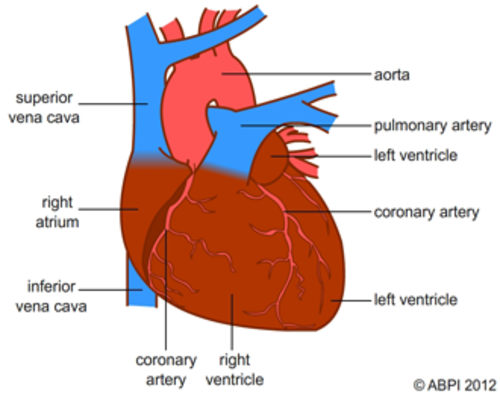
Anatomy of a Human Heart - uofmhealth Located between the lungs in the middle of the chest, the heart pumps blood through the network of arteries and veins known as the cardiovascular system. It pushes blood to the body's organs, tissues and cells. Blood delivers oxygen and nutrients to every cell and removes the carbon dioxide and other waste products made by those cells. Heart: illustrated anatomy - e-Anatomy - IMAIOS This interactive atlas of human heart anatomy is based on medical illustrations and cadaver photography. The user can show or hide the anatomical labels which provide a useful tool to create illustrations perfectly adapted for teaching. Anatomy of the heart: anatomical illustrations and structures, 3D model and photographs of dissection. Anatomy of the Heart - Medical Animation - YouTube This medical animation demonstrates the anatomy of the human heart, while explaining how the cardiovascular system functions. Explore more of our medical ani... PDF HEART - STRUCTURE - BiologyMad HEART - STRUCTURE • 4 sections Left atrium Right atrium Left ventricle Right ventricle • heart ry artery Pulmonary vein EAS the blood from he left hand side has to be pumped all around the body. • 2 lo heart Atrioventricular valves - between the atrium and the ventricles Semi-lunar valves - in the pulmonary artery and the aorta
Human Heart Pictures with Labels Inspirational File Heart Diagram Eng in 2020 | Human heart ...
Heart Diagram with Labels and Detailed Explanation - BYJUS Diagram of Heart. The human heart is the most crucial organ of the human body. It pumps blood from the heart to different parts of the body and back to the heart. The most common heart attack symptoms or warning signs are chest pain, breathlessness, nausea, sweating etc. The diagram of heart is beneficial for Class 10 and 12 and is frequently ...

Kenya Forensics Online Resource: CARDIAC MUSCLE TISSUE
19.1 Heart Anatomy - Anatomy and Physiology 2e | OpenStax Location of the Heart. The human heart is located within the thoracic cavity, medially between the lungs in the space known as the mediastinum. Figure 19.2 shows the position of the heart within the thoracic cavity. Within the mediastinum, the heart is separated from the other mediastinal structures by a tough membrane known as the pericardium, or pericardial sac, and sits in its own space ...
Label the heart - Teaching resources
How to Draw the Internal Structure of the Heart (with Pictures) To draw the internal structure of a human heart, follow the steps below. Part 1 Finding a Diagram 1 To find a good diagram, go to Google Images, and type in "The Internal Structure of the Human Heart". Find an image that displays the entire heart, and click on it to enlarge it. 2 Find a piece of paper and something to draw with.
Free Animal Cell Unlabeled, Download Free Clip Art, Free Clip Art on Clipart Library
Human Heart Diagram Labeled - Science Trends The heart's atrioventricular valves are structures that join the atria and ventricles of the heart together. This group of valves is comprised of the tricuspid valve and the mitral valve. Beyond this, there is a structure referred to as the aortic valve which separates the left ventricle and the aorta.
.png)
Parts Of The Heart - ProProfs Quiz
Human Heart Diagram Without Labels | Human heart diagram, Heart diagram ... ABOUT THIS ACTIVITY: Illustrates the pathway of blood through the heart. The areas of the heart with MORE oxygen are labeled with an "R". Students will color these areas RED. The areas of the heart with LESS oxygen are labeled with a "B". Students will color these areas BLUE.
Labeled picture of the heart – Graph Diagram
The Anatomy of the Heart, Its Structures, and Functions The heart is the organ that helps supply blood and oxygen to all parts of the body. It is divided by a partition (or septum) into two halves. The halves are, in turn, divided into four chambers. The heart is situated within the chest cavity and surrounded by a fluid-filled sac called the pericardium. This amazing muscle produces electrical ...
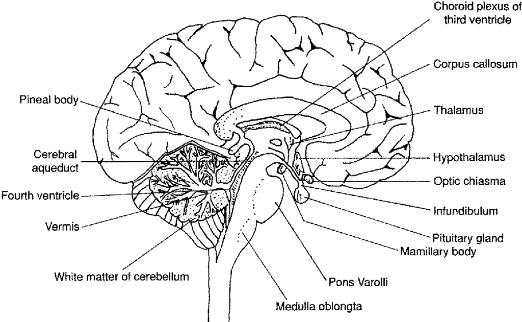
12 Best Images of Human Brain Diagram Worksheet - Human Brain Anatomy Coloring Page, Brain ...
Heart Blood Flow | Simple Anatomy Diagram, Cardiac Circulation ... - EZmed Step 2 involves the left atrium, the chamber of the heart that receives oxygenated blood from the lungs via the pulmonary veins. 3. Mitral Valve Step 3 involves the mitral valve. During diastole, when the heart is relaxed and filling with blood, the oxygenated blood from the left atrium will flow to the left ventricle.

Heart Anatomy: Labeled Diagram, Structures, Function, and Blood Flow Chambers of the Heart Let's begin with the chambers of the heart. There are 4 chambers, labeled 1-4 on the diagram below. To help simplify things, we can convert the heart into a square. We will then divide that square into 4 different boxes which will represent the 4 chambers of the heart.
Heart Diagram Labelling Activity
Heart Labeling Quiz: How Much You Know About Heart Labeling? Here is a Heart labeling quiz for you. The human heart is a vital organ for every human. The more healthy your heart is, the longer the chances you have of surviving, so you better take care of it. Take the following quiz to know how much you know about your heart. Questions and Answers 1. What is #1? 2. What is #2? 3. What is #3? 4. What is #4?
KS4 Biology The heart - Teaching resources
The structure of the heart - Structure and function of the heart ... It is located in the middle of the chest and slightly towards the left. The heart is a large muscular pump and is divided into two halves - the right-hand side and the left-hand side. The...

Inspirierend Heart Diagram With Labels Gcse
Structure of the Heart | SEER Training The human heart is a four-chambered muscular organ, shaped and sized roughly like a man's closed fist with two-thirds of the mass to the left of midline. The heart is enclosed in a pericardial sac that is lined with the parietal layers of a serous membrane. The visceral layer of the serous membrane forms the epicardium. Layers of the Heart Wall
Lab Heart Structure Labeling. - Heart Structure Labeling Lab Name Hannah Kingsley Date Time 11 ...
Heart: Anatomy and Function - Cleveland Clinic The parts of your heart are like the parts of a house. Your heart has: Walls. Chambers (rooms). Valves (doors). Blood vessels (plumbing). Electrical conduction system (electricity). Heart walls Your heart walls are the muscles that contract (squeeze) and relax to send blood throughout your body.

Business Diary: October 2011
WebMD - Better information. Better health. The heart is a muscular organ about the size of a fist, located just behind and slightly left of the breastbone. The heart pumps blood through the network of arteries and veins called the...
A labelled diagram of the heart - Document in A Level and IB Human Biology
A Labeled Diagram of the Human Heart You Really Need to See The human heart, comprises four chambers: right atrium, left atrium, right ventricle and left ventricle. The two upper chambers are called the left and the right atria, and the two lower chambers are known as the left and the right ventricles. The two atria and ventricles are separated from each other by a muscle wall called 'septum'.

Biology 156: Compendium Review Three -- Blood and everything related to it
Human Heart Diagram Without Labels - Labelling Worksheet The human heart is a muscle made up of four chambers, these are: Two upper chambers - the left atrium and right atrium Two lower chambers - the left and right ventricles. It's also made up of four valves - these are known as the tricuspid, pulmonary, mitral and aortic valves.
Heart Labelled | Teaching Resources
Post a Comment for "44 structure of the heart without labels"